


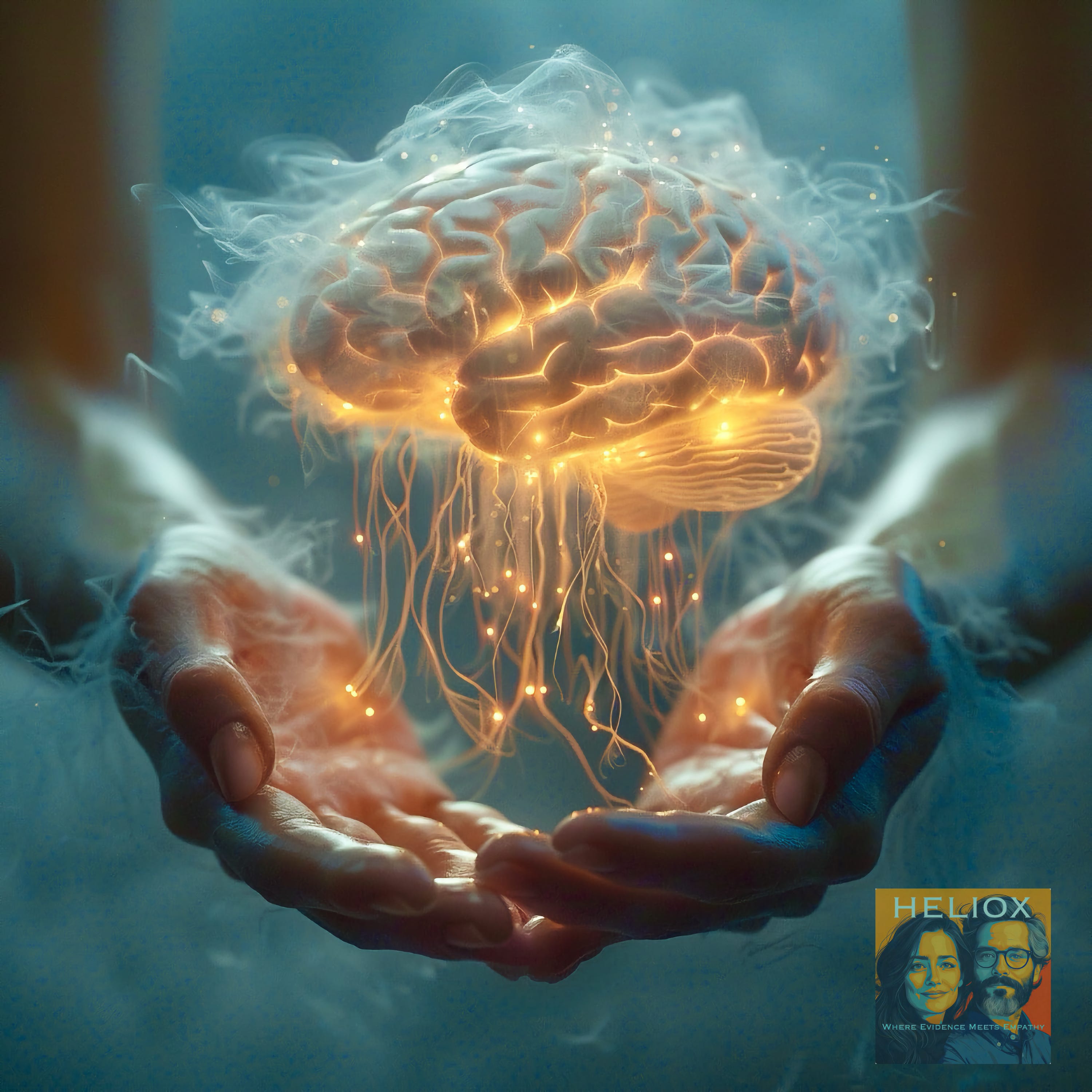
🧠 The Invisible Scars
What COVID is Really Doing to Our Brains

Haiku
Invisible scars
Reshape the mind's quiet paths—
Questions bloom in fog
With every article and podcast episode, we provide comprehensive study materials: References, Executive Summary, Briefing Document, Quiz, Essay Questions, Glossary, Timeline, Cast, FAQ, Table of Contents, Index, Polls, 3k Image, Fact Check, and
Comic at the very bottom of the page.
Essay
We're living through the largest uncontrolled experiment on human cognition in history, and most people don't even know they're subjects.
While the world moved on from pandemic panic to whatever fresh hell dominates this week's news cycle, researchers have been quietly documenting something that should terrify us all: COVID-19 is reshaping our brains in ways we're only beginning to understand.
The evidence is mounting, and it's not pretty.
The Brain Thief
Let's start with what we know. A major longitudinal study published in Nature—the kind of research that makes epidemiologists lose sleep—tracked 785 people between ages 51 and 81. Here's what makes this study gold standard: they had brain scans from before the pandemic even started.
Think about that. These researchers accidentally created the perfect control group for studying COVID's neurological effects. They could literally see what happened to people's brains after infection, comparing them to their own pre-pandemic baseline and to uninfected controls.
The results? Even mild COVID infections caused measurable brain changes. We're talking about physical alterations to gray matter—the brain tissue responsible for everything from memory to decision-making. The infected group showed greater reductions in gray matter thickness, particularly in areas linked to smell and memory processing.
But here's the kicker: these weren't just the people who ended up in ICUs. Most cases were mild to moderate. Only 15 participants needed hospitalization. The brain changes happened across the board.
The Aging Acceleration
The researchers put these changes in perspective using a metric that should make everyone pause: the level of brain tissue loss was equivalent to one to two extra years of normal aging.
One to two extra years. Per infection.
Let that sink in while you consider how many times you've been infected. Because if you think you've avoided COVID, you're probably wrong. The CDC estimates that by 2023, over 75% of Americans had been infected at least once. Many people have been infected multiple times.
Are we looking at a generation that's been neurologically aged by several years? The arithmetic is unsettling.
The Children We're Failing
Adults aren't the only ones paying the price. A study from Taiwan examined children before and after COVID infections, measuring behavioral, emotional, and social functioning. The results paint a disturbing picture of a generation whose emotional and social development has been derailed.
Children who'd been infected showed increased problems across multiple domains: school functioning, social interactions, behavioral regulation. They had more difficulty with attention, showed more oppositional behaviors, and demonstrated less active engagement with their parents.
The sex differences were particularly striking. Girls showed more widespread and severe impacts than boys—more aggression, anxiety, depression, social difficulties, and withdrawn behaviors. We're potentially watching the emergence of a generation of girls whose emotional and social development has been disrupted at a critical time.
The Mechanisms of Harm
How is this happening? The research points to several potential pathways, none of them reassuring.
One theory focuses on the olfactory system—the pathways that process smell. COVID's notorious ability to steal smell and taste may be just the tip of the iceberg. The virus appears to use these pathways as a highway into the brain, potentially causing a cascade of neurological damage that spreads beyond the initial infection site.
Another mechanism is neuroinflammation—the brain's immune response to infection. When your brain's immune system goes haywire, it can damage the very tissue it's trying to protect. This inflammation might persist long after the virus has been cleared from the body.
There's also the possibility that sensory deprivation itself—the loss of smell and taste—triggers brain changes. Use it or lose it applies to neural pathways too.
The Cognitive Casualties
The brain changes aren't just academic curiosities. They correlate with measurable cognitive decline, particularly in executive function and attention. The infected group in the UK study took significantly longer to complete tasks requiring mental flexibility and processing speed.
Translation: if you've felt like your brain isn't quite as sharp since COVID, you're not imagining it. The fog is real, and it's measurable.
This isn't just about feeling a little sluggish. Executive function governs our ability to plan, organize, switch between tasks, and control impulses. These are the cognitive skills that make us functional adults. When they're compromised, everything from work performance to relationship management becomes harder.
The Unanswered Questions
Here's what keeps researchers up at night: we don't know if these changes are permanent.
The brain has remarkable plasticity—its ability to form new connections and adapt to damage. But we're looking at population-level changes in brain structure and function. Even if individual brains can compensate, what does it mean when an entire generation shows measurable cognitive decline?
The most haunting question raised by this research is whether COVID infections might increase the risk of neurodegenerative diseases decades down the line. Are we looking at a future epidemic of dementia and Alzheimer's? The researchers can't answer that yet, but they're not ruling it out.
The Collective Denial
Despite mounting evidence, we're experiencing a form of collective denial about COVID's neurological effects. The dominant narrative has shifted to treating COVID as "just another respiratory virus," conveniently ignoring the growing body of research showing it's anything but.
This denial isn't accidental. It's psychologically necessary for a society that's decided to move on from pandemic precautions. Acknowledging the full scope of COVID's neurological impact would require admitting that our "return to normal" came at a devastating cost.
But reality doesn't care about our psychological comfort. The brain changes are happening whether we acknowledge them or not.
What This Means for All of Us
We're living through a public health disaster that's hiding in plain sight. While we argue about mask mandates and vaccine boosters, millions of people are experiencing subtle but measurable cognitive decline. Children are struggling with behavioral and emotional problems that may follow them into adulthood.
The implications ripple outward. If COVID is causing population-level cognitive decline, what does that mean for educational outcomes, workplace productivity, and social functioning? How do we support children whose emotional development has been disrupted? How do we plan for a future where neurodegenerative diseases might be more common?
These aren't abstract research questions. They're urgent policy challenges that we're collectively ignoring.
The Path Forward
The researchers are calling for longer follow-up studies, more detailed analysis of viral variants, and better understanding of who's most at risk. But research takes time, and people are being infected now.
What we need is a fundamental shift in how we think about COVID prevention. This isn't just about avoiding a few days of fever and cough. It's about protecting our brains and our children's developing minds from a virus that appears to cause lasting neurological damage.
The evidence is clear: COVID is changing our brains in ways we're only beginning to understand. The question isn't whether we can afford to take this seriously. It's whether we can afford not to.
Every infection is a roll of the dice with our most precious organ. And the house always wins.

Link References
"Mild COVID Linked to Brain Damage: What That Means for You" (Undated excerpt on brain therapies)
Episode Links
Youtube
Other Links to Heliox Podcast
YouTube
Substack
Podcast Providers
Spotify
Apple Podcasts
Patreon
FaceBook Group
STUDY MATERIALS
Briefing
Executive Summary
Recent research provides compelling evidence that SARS-CoV-2 infection, even in mild cases, is associated with significant and measurable changes in brain structure and cognitive function. These changes are particularly evident in brain regions related to olfaction and memory. Furthermore, a distinct body of research highlights an increase in behavioral, emotional, and social problems in children post-COVID-19 infection, suggesting broader neurological and developmental impacts beyond direct brain damage. While the full long-term implications are still being investigated, the findings underscore the need for continued monitoring and potential therapeutic interventions.
Main Themes and Key Findings
1. Structural Brain Changes Post-COVID-19 Infection
The study by Douaud et al. ("SARS-CoV-2 is associated with changes in brain structure in UK Biobank") provides the most robust evidence to date of brain structural changes linked to SARS-CoV-2 infection, utilizing a unique longitudinal design where participants were scanned both before and after infection.
Grey Matter Reduction: Infected individuals showed a "greater reduction in grey matter thickness and tissue contrast in the orbitofrontal cortex and parahippocampal gyrus." These areas are crucial for olfaction, memory, and executive functions.
Tissue Damage Markers: There were "greater changes in markers of tissue damage in regions that are functionally connected to the primary olfactory cortex." This suggests damage spreading through olfactory pathways.
Global Brain Size Reduction: SARS-CoV-2 cases exhibited a "greater reduction in global brain size." This indicates a diffuse loss of grey matter.
Impact on Mild Cases: Crucially, these structural and cognitive effects were observed even after excluding patients who had been hospitalized, meaning "the impact of SARS-CoV-2 infection can be detected in milder cases."
Olfactory Pathway Involvement: The consistent clinical feature of disturbed olfaction and gustation in COVID-19 patients is a potential mechanism for these brain changes. The study notes that "Such loss of sensory olfactory inputs to the brain could lead to a loss of grey matter in olfactory-related brain regions."
Comparison to Normal Aging: The observed changes, although modest in percentage (e.g., 1.3% to 1.8% for parahippocampal/entorhinal cortex volume), are significant when compared to normal age-related atrophy, where hippocampal volume loss is around "0.2% (in middle age) to 0.3% (in older age) per year."
Possible Mechanisms: The authors suggest that these changes could be "the in vivo hallmarks of a degenerative spread of the disease through olfactory pathways, of neuroinflammatory events, or of the loss of sensory input due to anosmia."
Long-Term Implications: The study raises the concern that "longer-term consequences of SARS-CoV-2 infection might in time contribute to Alzheimer’s disease or other forms of dementia."
2. Cognitive Decline and Executive Function Impairment
Beyond structural changes, SARS-CoV-2 infection has been linked to measurable cognitive impacts.
Cognitive Decline: Douaud et al. found a "greater cognitive decline between the two time points" in infected participants. This was still observed even when excluding hospitalized patients.
Executive Function Worsening: Specifically, infected individuals showed "a significantly greater increase in the time taken to complete trails A (numeric) and B (alphanumeric) of the Trail Making Test." These tests are "known to be sensitive to detect impairment of executive function and attention."
Association with Cerebellum: The duration to complete the alphanumeric trail B was "associated post hoc with the longitudinal changes in the cognitive part of the cerebellum, namely crus II," which is also involved in olfactory tasks.
"Brain Fog": Ziyad Al-Aly's excerpt ("Mounting research shows that COVID-19 leaves its mark on the brain...") directly references "Brain fog" as a cognitive health issue linked to COVID-19, aligning with the observed impairments in memory, attention, and executive functioning.
3. Increased Behavioral, Emotional, and Social Problems in Children
The Taiwanese study ("Increased post-COVID-19 behavioral, emotional, and social problems in Taiwanese children") provides crucial insights into the post-infection mental health of children.
Study Design: This longitudinal study compared 84 children aged 6-16 with SARS-CoV-2 infection to 84 age-, sex-, and IQ-matched controls, assessing them 1-6 months post-infection. They used comprehensive tools like SNAP-IV for ADHD symptoms, CBCL for behavioral/emotional problems, and SAICA for social adjustment.
Significant Increases Across Multiple Domains: Children who had COVID-19 showed statistically significant increases in various behavioral, emotional, and social problems compared to controls and even compared to their own pre-COVID condition (where data was available).
ADHD-related symptoms: Increased "Inattention" and "Opposition" were significantly higher post-COVID.
Behavioral/Emotional Problems (CBCL): Significant increases were noted in:
"Aggressive behavior"
"Anxious/Depressed" symptoms
"Attention problems"
"Delinquent behavior"
"Social problems"
"Somatic complaints" (showing a progressive increase: Control < Pre-COVID < Post-COVID)
"Thought problems"
"Withdrawn" symptoms
"Internalizing problems" and "Externalizing problems" (both broad-band dimensions showed significant increases).
Social Adjustment Problems (SAICA): Significant impacts were observed on:
"Overall school functions" (Control < Pre-COVID < Post-COVID)
"Academic performance"
"School attitude"
"Social interaction"
"School behavioral problem"
"Problem with parents"
"Interaction with parents"
Specific Severity: For "Somatic complaints" and "Overall school functions," the issues were significantly worse post-COVID compared to both pre-COVID and control groups, indicating a direct worsening effect after infection.
Pre-existing Conditions: While 16.7% of the COVID-infected group had an ADHD diagnosis, the study design aimed to control for major pre-existing psychiatric or neurological disorders. The longitudinal comparison to "Pre-COVID condition" further strengthens the link to the infection.
4. Therapeutic Approaches for Post-COVID Brain Damage
An undated excerpt titled "Mild COVID Linked to Brain Damage: What That Means for You" briefly outlines potential therapeutic interventions.
Multi-Disciplinary Therapies: It suggests a range of therapies, including "Neuromuscular therapy, Sensorimotor therapy, Cognitive therapy, Occupational therapy, Vision therapy, Vestibular therapy, Psychotherapy, And more."
Cognitive Therapy Focus: Specifically, cognitive therapy aims to "address any cognitive impairments, including issues related to memory, attention, executive functioning, and word finding." Examples include "word games, puzzles, finding hidden patterns and memorizing a list of words."
Role of Cardio Exercise: The source also notes that "Preparing the brain with cardio exercise makes it more receptive to the other therapy appointments you have each day."
Conclusion
The evidence from these sources strongly suggests that SARS-CoV-2 infection can lead to measurable and significant neurological and neurodevelopmental consequences. In adults, even mild cases are associated with structural brain changes, particularly in olfactory and limbic regions, and cognitive decline affecting executive functions. In children, there is a marked increase in a wide array of behavioral, emotional, and social problems post-infection, impacting daily functioning and school performance. While the long-term reversibility of these effects is still under investigation, the findings highlight a critical need for awareness, early identification, and comprehensive therapeutic interventions to mitigate the lasting impact of COVID-19 on brain health across all age groups.
Quiz & Answer Key
Instructions: Answer each question in 2-3 sentences.
What was the primary objective of the Taiwanese study ("Increased post-COVID-19 behavioral, emotional, and social problems in Taiwanese children.pdf") regarding children infected with SARS-CoV-2?
Describe the demographic characteristics of the participants in the Taiwanese study. How were the COVID group and control group matched?
Name three specific measurement tools used in the Taiwanese study to assess behavioral, emotional, and social problems in children.
According to the Taiwanese study, what were some of the significant behavioral/emotional problems observed in the post-COVID condition compared to the control group, as measured by the CBCL?
What key findings did the "SARS-CoV-2 is associated with changes in brain structure in UK Biobank" study report regarding brain structure changes in individuals who had mild COVID-19?
How did the UK Biobank study ensure that the observed brain changes were likely due to SARS-CoV-2 infection rather than pre-existing conditions?
Which specific brain regions were most consistently affected by SARS-CoV-2 infection, according to the UK Biobank study, and what sensory system are these regions primarily associated with?
Besides structural brain changes, what cognitive decline was significantly observed in the SARS-CoV-2-positive group in the UK Biobank study, even in non-hospitalized cases?
According to the "Mild COVID Linked to Brain Damage" source, what types of therapies are suggested for addressing cognitive impairments associated with mild COVID?
What long-term implications, particularly concerning neurodegenerative diseases, are raised by the findings of the UK Biobank study regarding brain changes post-COVID-19?
Quiz Answer Key
The primary objective of the Taiwanese study was to investigate the prevalence of behavioral, emotional, and social problems in children aged 6-16 who had been infected with SARS-CoV-2, comparing them to a matched control group. The study aimed to identify any increased issues in post-COVID conditions.
The participants in the Taiwanese study included 84 children with SARS-CoV-2 infection and 84 non-infected healthy controls. Both groups were carefully matched by age, sex, and Full-Scale IQ (FIQ), ensuring comparability between the infected and control cohorts.
Three specific measurement tools used in the Taiwanese study were the Kiddie-epidemiologic version of the Schedule for Affective Disorders and Schizophrenia (K-SADS-E) for psychiatric diagnoses, the Chinese version of the Swanson, Nolan, and Pelham, version IV scale (SNAP-IV) for ADHD-related symptoms, and the Child Behavior Checklist (CBCL) for a broad range of behavioral/emotional problems.
According to the Taiwanese study, as measured by the CBCL, significant behavioral/emotional problems observed in the post-COVID condition included increased aggressive behavior, anxious/depressed symptoms, attention problems, delinquent behavior, social problems, somatic complaints, thought problems, and withdrawn symptoms, compared to the control group. Internalizing and externalizing problems were also significantly higher.
The UK Biobank study reported a greater reduction in grey matter thickness and tissue contrast in the orbitofrontal cortex and parahippocampal gyrus, increased tissue damage in regions connected to the primary olfactory cortex, and a greater reduction in global brain size in SARS-CoV-2 cases. These effects were seen even in milder cases.
The UK Biobank study utilized a longitudinal design where participants had undergone brain imaging before being infected with SARS-CoV-2 and were re-imaged after infection. This pre-infection baseline data allowed researchers to compare individual changes over time, reducing the likelihood of misinterpreting pre-existing risk factors as disease effects.
The brain regions most consistently affected by SARS-CoV-2 infection were primarily limbic and olfactory cortical systems. These included the orbitofrontal cortex, parahippocampal gyrus, anterior cingulate cortex, insula, ventral striatum, amygdala, and hippocampus, all of which are functionally connected to the primary olfactory cortex.
The SARS-CoV-2-positive group in the UK Biobank study showed a significantly greater cognitive decline in executive function. Specifically, they took a significantly longer time to complete trails A and B of the Trail Making Test, even after excluding the few hospitalized cases.
For addressing cognitive impairments related to mild COVID, the "Mild COVID Linked to Brain Damage" source suggests various therapies. These include neuromuscular therapy, sensorimotor therapy, cognitive therapy (e.g., word games, puzzles, memorizing lists), occupational therapy, vision therapy, vestibular therapy, and psychotherapy.
The UK Biobank study raises the possibility that the observed long-term structural brain changes, particularly in memory-related regions like the parahippocampal gyrus, entorhinal cortex, and hippocampus, might in time contribute to the risk of developing Alzheimer's disease or other forms of dementia. This has led to the formation of international consortia to investigate this link further.
Essay Questions
Compare and contrast the methodologies, participant recruitment, and findings of the Taiwanese study on children and the UK Biobank study on adults. Discuss how the different cohorts and research designs contribute to our understanding of COVID-19's impact on the brain and behavior.
Discuss the evidence presented across the sources that suggests a connection between olfactory system dysfunction and broader neurological and cognitive impairments post-COVID-19. How might this connection explain some of the observed brain changes and cognitive declines?
Analyze the immediate and potential long-term implications of SARS-CoV-2 infection on cognitive function and brain health as highlighted by the UK Biobank study. What are the key limitations of the study in predicting long-term outcomes, and what future research is suggested?
Based on the provided texts, what are the different types of problems—behavioral, emotional, social, and cognitive—that have been linked to COVID-19 infection in both children and adults? Discuss the potential mechanisms underlying these diverse impacts.
Evaluate the significance of longitudinal study designs, as exemplified by the UK Biobank study, in understanding the effects of diseases like COVID-19. How do such designs strengthen the conclusions drawn compared to cross-sectional studies, and what specific advantages did the UK Biobank's pre-infection imaging data offer?
Glossary of KeyTerms
ADHD-related symptoms: Symptoms associated with Attention-Deficit/Hyperactivity Disorder, including inattention, hyperactivity, and impulsivity. Measured by tools like SNAP-IV.
Anosmia: Complete loss of the sense of smell.
Anterograde degeneration: Degeneration that proceeds from the cell body along the axon to the synapse, often occurring after an injury to the neuron's cell body or axon.
Anxious/Depressed symptoms: Behavioral and emotional problems characterized by feelings of anxiety, sadness, worry, and low mood. A subscale of the CBCL.
Attention problems: Difficulties with sustaining focus, concentrating, and being easily distracted. A subscale of the CBCL.
CBCL (Child Behavior Checklist): A parent-rated questionnaire used to assess a wide range of behavioral and emotional problems in children and adolescents, yielding narrow-band syndromes and broad-band dimensions (Internalizing and Externalizing problems).
Cognitive Decline: A measurable decrease in cognitive functions such as memory, attention, executive functioning, and processing speed, observed over time.
Cortical Thickness: A measure of the thickness of the cerebral cortex, which is the outer layer of the brain responsible for higher-level functions. Reductions can indicate atrophy.
Crus II: A specific lobule in the cerebellum, known to be involved in cognitive functions, including those related to olfaction.
Cerebrospinal Fluid (CSF) Volume: The total volume of the fluid surrounding the brain and spinal cord. An increase in CSF volume in the context of brain atrophy can suggest a diffuse loss of grey matter.
Diffusion Indices (e.g., orientation dispersion, isotropic volume fraction, mean diffusivity): Measures derived from diffusion MRI that provide insights into tissue microstructure integrity. Changes can indicate tissue damage or alterations.
DSM-5: The fifth edition of the Diagnostic and Statistical Manual of Mental Disorders, published by the American Psychiatric Association, used for the classification and diagnosis of mental disorders.
Executive Function: A set of cognitive processes that are necessary for the cognitive control of behavior, including attention, working memory, planning, problem-solving, and inhibition.
Externalizing problems: A broad-band dimension of the CBCL, typically including delinquent behavior and aggressive behavior, reflecting outward-directed problematic behaviors.
Full-Scale IQ (FIQ): A composite score derived from the Wechsler Intelligence Scale for Children (WISC-V) that represents a child's overall intellectual ability.
Grey Matter Volume/Thickness: The volume or thickness of brain tissue composed primarily of neuronal cell bodies, dendrites, and unmyelinated axons. Reductions can indicate neuronal loss or atrophy.
Gustation/Hypogeusia: The sense of taste; hypogeusia is a reduced ability to taste.
Hippocampus: A brain region located in the medial temporal lobe, crucial for memory formation, particularly episodic memory.
Hyposmia: A reduced ability to smell.
IDPs (Imaging-Derived Phenotypes): Summary measures obtained from brain imaging data, each describing a different aspect of brain structure or function (e.g., grey matter volume, cortical thickness, diffusion measures).
Insula: A brain region deep within the lateral sulcus, involved in various functions including taste (primary gustatory cortex), emotion, and interoception; directly connected to the primary olfactory cortex.
Internalizing problems: A broad-band dimension of the CBCL, typically including anxious/depressed symptoms, withdrawn symptoms, and somatic complaints, reflecting inward-directed problematic behaviors.
K-SADS-E (Kiddie-epidemiologic version of the Schedule for Affective Disorders and Schizophrenia): An investigator-rated semi-structured interview scale used for the diagnosis of psychiatric disorders according to DSM-5 criteria.
Limbic System: A complex set of brain structures that includes the hippocampus, amygdala, and parts of the cortex, involved in emotion, motivation, memory, and olfaction.
Longitudinal Study: A research design that involves repeated observations of the same variables (e.g., participants) over short or long periods of time.
Mean Diffusivity: A diffusion MRI measure that quantifies the average magnitude of water diffusion within tissue. Increased mean diffusivity can indicate tissue damage, such as loss of cellular integrity or edema.
MRI (Magnetic Resonance Imaging): A non-invasive imaging technique used to visualize internal structures of the body, including the brain, using strong magnetic fields and radio waves.
Multi-system inflammatory syndrome in children (MIS-C): A serious condition associated with COVID-19 where different body parts can become inflamed, including the heart, lungs, kidneys, brain, skin, eyes, or gastrointestinal organs.
Neuromuscular therapy: A type of manual therapy focused on treating soft tissue pain and dysfunction.
Olfactory Bulb: A neural structure of the vertebrate forebrain involved in the sense of smell, receiving direct input from olfactory receptor neurons.
Olfactory Cortex (Primary/Secondary): Regions of the brain responsible for processing olfactory (smell) information. The piriform cortex is considered primary, while the orbitofrontal cortex is often referred to as secondary.
Omicron variants: A highly transmissible variant of SARS-CoV-2 that caused a significant wave of COVID-19 infections.
Orbitofrontal Cortex: A prefrontal cortex region located at the front of the brain, involved in decision-making, reward processing, and olfaction (often called the secondary olfactory cortex).
Parahippocampal Gyrus: A cortical region surrounding the hippocampus, part of the limbic system, involved in memory encoding and retrieval, and directly connected to the piriform and entorhinal cortices.
Piriform Cortex: The primary olfactory cortex in the brain, receiving direct input from the olfactory bulb.
Psychotherapy: A range of therapeutic techniques used to improve mental health and well-being, often involving talking to a therapist.
Rapid Antigen Test Kit: A diagnostic test used to detect the presence of SARS-CoV-2 viral antigens in a respiratory sample, providing quick results.
SARS-CoV-2: The severe acute respiratory syndrome coronavirus 2, which is the virus that causes COVID-19.
SAICA (Social Adjustment Inventory for Children and Adolescents): A measurement tool used to assess a child's social adjustment, including school functions, peer relationships, and home behaviors.
Sensorimotor therapy: Therapy that focuses on integrating sensory information with motor responses to improve movement and function.
SNAP-IV (Swanson, Nolan, and Pelham, version IV scale): A 26-item parent-rated scale designed to measure symptoms of inattention, hyperactivity/impulsivity, and opposition, commonly used for ADHD-related symptoms.
Somatic complaints: Physical symptoms that are medically unexplained but are often linked to psychological factors, such as headaches or stomachaches. A subscale of the CBCL.
Thought problems: Difficulties with coherent thought processes, including obsessive thoughts or unusual thinking. A subscale of the CBCL.
Trail Making Test (Parts A & B): A neuropsychological test of visual attention and task switching, used to assess executive function and cognitive processing speed.
UK Biobank: A large-scale biomedical database and research resource, containing in-depth genetic and health information from half a million UK participants, including brain imaging data.
Vertex-wise/Voxel-wise analysis: Methods of brain imaging analysis that examine changes at individual points (vertices on a surface, voxels in a volume) across the brain, allowing for detailed spatial mapping of effects.
Withdrawn symptoms: Behaviors characterized by social isolation, shyness, and a lack of engagement with others. A subscale of the CBCL.
Timeline of Main Events
Pre-COVID-19 Pandemic (Dates unspecified, prior to March 2020 and April 2022)
UK Biobank Participants' First Scans: Thousands of UK Biobank participants, aged 45 and over, received initial brain scans using MRI, forming a crucial baseline for future longitudinal studies.
Taiwanese Children Control Group Established: A historical control group of 80 non-infected healthy children was identified in Taiwan for a mental health study. These children did not experience the COVID-19 pandemic.
Pre-existing Mental Health Data: The Chinese version of the K-SADS-E, SNAP-IV, CBCL, and SAICA scales were established and widely used in clinical and epidemiological studies on ADHD and other behavioral/emotional problems in children in Taiwan.
March 2020 - April 2021
SARS-CoV-2 Infection Period (UK Biobank Study): 401 UK Biobank participants tested positive for SARS-CoV-2 infection between their two MRI scans. The specific strains of the virus during this period included the original strain and variants of concern present in the UK from October 2020 onwards (predominantly Alpha, possibly Beta and Gamma). Very few, if any, were infected with the Delta variant, which appeared in the UK in April 2021.
February - May 2021
UK Biobank Participants' Second Scans: The second brain scans for the SARS-CoV-2-positive participants in the UK Biobank study were acquired. On average, 141 days separated their diagnosis and this second scan.
March 7, 2022
"SARS-CoV-2 is associated with changes in brain structure in UK Biobank" Published: This open-access article, co-authored by Gwenaëlle Douaud, Stephen M. Smith, and others, was published in Nature, detailing the findings of the longitudinal brain imaging study of SARS-CoV-2.
April 2022
Omicron Variant Outbreak in Taiwan: The outbreak of the COVID-19 pandemic began with the spread of Omicron variants in Taiwan. This marks a significant shift in the pandemic's impact on children in the region.
Additional Taiwanese Children Controls Enrolled: 4 controls without SARS-CoV-2 infection were enrolled after April 2022 for the Taiwanese children's mental health study, confirmed by parental reports.
October 6, 2022 - April 30, 2023
Taiwanese Children COVID-19 Group Clinical Assessments: 84 children aged 6–16 with SARS-CoV-2 infection underwent clinical assessments 1–6 months after testing positive for COVID-19 (mean duration, 163 ± 55 days) at National Taiwan University Children’s Hospital.
2024
"Post COVID-19 condition and behavioral manifestations in Taiwanese children" Publication: Li CJ, Yu HR, Kou KC, et al. published their findings in Pediatric International.
September 2: The manuscript "Increased post-COVID-19 behavioral, emotional, and social problems in Taiwanese children" was received.
October 10: The manuscript "Increased post-COVID-19 behavioral, emotional, and social problems in Taiwanese children" was received in revised form.
October 16: The manuscript "Increased post-COVID-19 behavioral, emotional, and social problems in Taiwanese children" was accepted.
2025
Publication of "Increased post-COVID-19 behavioral, emotional, and social problems in Taiwanese children": This article, authored by C.-Y. Shang, S.S.-F. Gau, and others, is slated for publication in Journal of the Formosan Medical Association 124.
Cast of Characters
Researchers/Authors:
Gwenaëlle Douaud: Co-first author of "SARS-CoV-2 is associated with changes in brain structure in UK Biobank," affiliated with FMRIB Centre, Wellcome Centre for Integrative Neuroimaging (WIN), Nuffield Department of Clinical Neurosciences, University of Oxford. She conceived the brain imaging study and interpreted the results.
Stephen M. Smith: Co-first author of "SARS-CoV-2 is associated with changes in brain structure in UK Biobank," affiliated with FMRIB Centre, Wellcome Centre for Integrative Neuroimaging (WIN), Nuffield Department of Clinical Neurosciences, University of Oxford. He carried out the imaging analyses and co-wrote the paper.
Ziyad Al-Aly: Author of "Mounting research shows that COVID-19 leaves its mark on the brain, including significant drops in I.pdf". He is the Chief of Research and Development at VA St. Louis Health Care System and a Clinical Epidemiologist at Washington University in St. Louis.
Shih-Fen S. Gau (S.S.-F. Gau): Corresponding author of "Increased post-COVID-19 behavioral, emotional, and social problems in Taiwanese children," affiliated with the Department of Psychiatry, National Taiwan University Hospital and College of Medicine, Taipei, Taiwan. A prolific researcher on mental disorders in children in Taiwan, especially ADHD.
Chih-Ying Shang (C.-Y. Shang): An author on "Increased post-COVID-19 behavioral, emotional, and social problems in Taiwanese children." Also affiliated with the Department of Psychiatry, National Taiwan University Hospital and College of Medicine, Taipei, Taiwan.
Soojin Lee: Author on "SARS-CoV-2 is associated with changes in brain structure in UK Biobank," affiliated with FMRIB Centre, Wellcome Centre for Integrative Neuroimaging (WIN), Nuffield Department of Clinical Neurosciences, University of Oxford.
Fidel Alfaro-Almagro: Author on "SARS-CoV-2 is associated with changes in brain structure in UK Biobank," affiliated with FMRIB Centre, Wellcome Centre for Integrative Neuroimaging (WIN), Nuffield Department of Clinical Neurosciences, University of Oxford.
Christoph Arthofer: Author on "SARS-CoV-2 is associated with changes in brain structure in UK Biobank," affiliated with FMRIB Centre, Wellwellcome Centre for Integrative Neuroimaging (WIN), Nuffield Department of Clinical Neurosciences, University of Oxford.
Chaoyue Wang: Author on "SARS-CoV-2 is associated with changes in brain structure in UK Biobank," affiliated with FMRIB Centre, Wellcome Centre for Integrative Neuroimaging (WIN), Nuffield Department of Clinical Neurosciences, University of Oxford.
Paul McCarthy: Author on "SARS-CoV-2 is associated with changes in brain structure in UK Biobank," affiliated with FMRIB Centre, Wellcome Centre for Integrative Neuroimaging (WIN), Nuffield Department of Clinical Neurosciences, University of Oxford.
Frederik Lange: Author on "SARS-CoV-2 is associated with changes in brain structure in UK Biobank," affiliated with FMRIB Centre, Wellcome Centre for Integrative Neuroimaging (WIN), Nuffield Department of Clinical Neurosciences, University of Oxford.
Jesper L. R. Andersson: Author on "SARS-CoV-2 is associated with changes in brain structure in UK Biobank," affiliated with FMRIB Centre, Wellcome Centre for Integrative Neuroimaging (WIN), Nuffield Department of Clinical Neurosciences, University of Oxford.
Ludovica Griffanti: Author on "SARS-CoV-2 is associated with changes in brain structure in UK Biobank," affiliated with FMRIB Centre, Wellcome Centre for Integrative Neuroimaging (WIN), Nuffield Department of Clinical Neurosciences, University of Oxford, and OHBA, Wellcome Centre for Integrative Neuroimaging (WIN), Department of Psychiatry, University of Oxford.
Eugene Duff: Author on "SARS-CoV-2 is associated with changes in brain structure in UK Biobank," affiliated with FMRIB Centre, Wellcome Centre for Integrative Neuroimaging (WIN), Nuffield Department of Clinical Neurosciences, University of Oxford, and Department of Paediatrics, University of Oxford.
Saad Jbabdi: Author on "SARS-CoV-2 is associated with changes in brain structure in UK Biobank," affiliated with FMRIB Centre, Wellcome Centre for Integrative Neuroimaging (WIN), Nuffield Department of Clinical Neurosciences, University of Oxford.
Bernd Taschler: Author on "SARS-CoV-2 is associated with changes in brain structure in UK Biobank," affiliated with FMRIB Centre, Wellcome Centre for Integrative Neuroimaging (WIN), Nuffield Department of Clinical Neurosciences, University of Oxford. He co-supervised the statistical analyses.
Peter Keating: Author on "SARS-CoV-2 is associated with changes in brain structure in UK Biobank," affiliated with Ear Institute, University College London. He contributed to conceiving the brain imaging study.
Anderson M. Winkler: Author on "SARS-CoV-2 is associated with changes in brain structure in UK Biobank," affiliated with National Institute of Mental Health, National Institutes of Health, Bethesda, MD, USA. He co-supervised the statistical analyses.
Rory Collins: Author on "SARS-CoV-2 is associated with changes in brain structure in UK Biobank," affiliated with Nuffield Department of Population Health, University of Oxford. He is the chief executive and principal investigator of UK Biobank and contributed to the creation of the UK Biobank COVID-19 re-imaging project.
Paul M. Matthews: Author on "SARS-CoV-2 is associated with changes in brain structure in UK Biobank," affiliated with UK Dementia Research Institute and Department of Brain Sciences, Imperial College, London. He contributed to the creation of the UK Biobank COVID-19 re-imaging project.
Naomi Allen: Author on "SARS-CoV-2 is associated with changes in brain structure in UK Biobank," affiliated with Nuffield Department of Population Health, University of Oxford. She is the chief scientist for UK Biobank and contributed to the creation of the UK Biobank COVID-19 re-imaging project.
Karla L. Miller: Author on "SARS-CoV-2 is associated with changes in brain structure in UK Biobank," affiliated with FMRIB Centre, Wellcome Centre for Integrative Neuroimaging (WIN), Nuffield Department of Clinical Neurosciences, University of Oxford. She contributed to conceiving the brain imaging study and creating the UK Biobank COVID-19 re-imaging project.
Thomas E. Nichols: Author on "SARS-CoV-2 is associated with changes in brain structure in UK Biobank," affiliated with Big Data Institute, University of Oxford. He co-supervised the statistical analyses.
Li CJ: Author on "Post COVID-19 condition and behavioral manifestations in Taiwanese children."
Yu HR: Author on "Post COVID-19 condition and behavioral manifestations in Taiwanese children."
Kou KC: Author on "Post COVID-19 condition and behavioral manifestations in Taiwanese children."
Chen SH: Author on "Differential clinical characteristics and performance of home antigen tests between parents and children after household transmission of SARS-CoV-2 during the Omicron variant pandemic."
Wu JL: Author on "Differential clinical characteristics and performance of home antigen tests between parents and children after household transmission of SARS-CoV-2 during the Omicron variant pandemic."
Liu YC: Author on "Differential clinical characteristics and performance of home antigen tests between parents and children after household transmission of SARS-CoV-2 during the Omicron variant pandemic."
Chen YL: Author on "The Mandarin version of the Kiddie-Schedule for Affective Disorders and Schizophrenia-Epidemiological version for DSM-5 - a psychometric study" and "Prevalence of DSM-5 mental disorders in a nationally representative sample of children in Taiwan: methodology and main findings."
Shen LJ: Author on "The Mandarin version of the Kiddie-Schedule for Affective Disorders and Schizophrenia-Epidemiological version for DSM-5 - a psychometric study."
Chong MY: Author on "A 3-year panel study of mental disorders among adolescents in Taiwan."
Chen TH: Author on "A 3-year panel study of mental disorders among adolescents in Taiwan."
Lin YJ: Author on "Psychiatric comorbidity and social adjustment difficulties in children with disruptive mood dysregulation disorder: a national epidemiological study."
Tseng WL: Author on "Psychiatric comorbidity and social adjustment difficulties in children with disruptive mood dysregulation disorder: a national epidemiological study."
Chang JC: Author on "Distinct developmental changes in regional gray matter volume and covariance in individuals with attention-deficit hyperactivity disorder: a longitudinal voxel-based morphometry study."
Lin HY: Author on "Distinct developmental changes in regional gray matter volume and covariance in individuals with attention-deficit hyperactivity disorder: a longitudinal voxel-based morphometry study."
Kuo RN: Author on "Burden of mental disorders in children in the general population and in health facilities: discrepancies in years lived with disability based on national prevalence estimates between populations receiving care or not."
Chiang HL: Author on "Shared intrinsic functional connectivity alterations as a familial risk marker for ADHD: a resting-state functional magnetic resonance imaging study with sibling design."
Tseng WI: Author on "Shared intrinsic functional connectivity alterations as a familial risk marker for ADHD: a resting-state functional magnetic resonance imaging study with sibling design."
Wey HY: Author on "Shared intrinsic functional connectivity alterations as a familial risk marker for ADHD: a resting-state functional magnetic resonance imaging study with sibling design."
Chiu YN: Author on "Developmental changes of autistic symptoms, ADHD symptoms, and attentional performance in children and adolescents with autism spectrum disorder."
Wu YY: Author on "Developmental changes of autistic symptoms, ADHD symptoms, and attentional performance in children and adolescents with autism spectrum disorder."
Tung YH: Author on "Whole brain white matter tract deviation and idiosyncrasy from normative development in autism and ADHD and unaffected siblings link with dimensions of psychopathology and cognition."
Chen CL: Author on "Whole brain white matter tract deviation and idiosyncrasy from normative development in autism and ADHD and unaffected siblings link with dimensions of psychopathology and cognition."
Swanson JM: Original developer of the SNAP-IV scale.
Kraemer HC: Contributor to research on the clinical relevance of MTA findings using SNAP-IV.
Hinshaw SP: Contributor to research on the clinical relevance of MTA findings using SNAP-IV.
John K: Author of the Social Adjustment Inventory for Children and Adolescents (SAICA).
Gammon GD: Author of the Social Adjustment Inventory for Children and Adolescents (SAICA).
Prusoff BA: Author of the Social Adjustment Inventory for Children and Adolescents (SAICA).
Soong WT: Author on research regarding methylphenidate in children with ADHD in Taiwan.
Organizations/Institutions:
National Taiwan University Hospital and College of Medicine, Taipei, Taiwan: Key institution for the "Increased post-COVID-19 behavioral, emotional, and social problems in Taiwanese children" study.
National Taiwan University Children’s Hospital, Taipei, Taiwan: Where clinical assessments for the Taiwanese children study were conducted.
UK Biobank: A large-scale biomedical database and research resource that played a central role in the "SARS-CoV-2 is associated with changes in brain structure in UK Biobank" study.
FMRIB Centre, Wellcome Centre for Integrative Neuroimaging (WIN), Nuffield Department of Clinical Neurosciences, University of Oxford, UK: Primary affiliation for many authors of the UK Biobank brain study.
Washington University in St. Louis: Affiliation for Ziyad Al-Aly.
VA St. Louis Health Care System: Affiliation for Ziyad Al-Aly.
Public Health England (PHE), Public Health Scotland (PHS), Secure Anonymised Information Linkage (SAIL, Wales): Provided diagnostic antigen test results data to UK Biobank.
Other Entities:
Children (6-16 years old) with SARS-CoV-2 infection (Taiwanese Study): 84 participants who tested positive for COVID-19, undergoing clinical assessments.
Healthy Control Children (Taiwanese Study): 84 participants matched by age, sex, and IQ, with no history of SARS-CoV-2 infection or major psychiatric/neurological disorders.
UK Biobank Participants (51-81 years old) with SARS-CoV-2 infection: 401 individuals who tested positive for the virus between their two brain scans. This group includes 15 who were hospitalized, 2 of whom received critical care.
UK Biobank Control Participants (51-81 years old): 384 individuals matched for age, sex, ethnicity, and time elapsed between scans, who tested negative for SARS-CoV-2 or had no medical record of COVID-19.
FAQ
1. What are the observed behavioral, emotional, and social problems in children after COVID-19 infection?
A study conducted at National Taiwan University Children’s Hospital found increased post-COVID-19 behavioral, emotional, and social problems in children aged 6–16. Compared to a control group, children who had contracted SARS-CoV-2 showed significantly higher scores in several areas according to parent-rated questionnaires:
Behavioral/Emotional Problems (CBCL): Increased aggressive behavior, anxious/depressed symptoms, attention problems, delinquent behavior, social problems, somatic complaints, thought problems, and withdrawn symptoms. Both internalizing (anxious/depressed, withdrawn, somatic complaints) and externalizing (delinquent, aggressive) problem scores were significantly higher.
ADHD Symptomatology (SNAP-IV): A notable increase in inattention and opposition symptoms. Hyperactivity/impulsivity also showed an increase, though not statistically significant across all comparisons.
Social Adjustment (SAICA): Significant difficulties in overall school functions, academic performance, school attitude, social interaction, school behavioral problems, and relationships/interaction with parents.
These issues were observed even in children who did not have a prior history of major psychiatric disorders.
2. How does COVID-19 infection affect brain structure and cognitive function in adults, particularly in milder cases?
Research, including a longitudinal imaging study using UK Biobank data, indicates that SARS-CoV-2 infection is associated with detectable changes in brain structure and cognitive decline, even in individuals with milder cases of COVID-19. Key findings include:
Grey Matter Reduction: A greater reduction in grey matter thickness and tissue contrast was observed in specific brain regions, notably the orbitofrontal cortex and parahippocampal gyrus. These regions are functionally connected to the primary olfactory cortex and are involved in memory and executive functions.
Tissue Damage Markers: Increased markers of tissue damage were found in regions functionally connected to the primary olfactory cortex.
Global Brain Size Reduction: A greater overall reduction in global brain size was noted in SARS-CoV-2 cases.
Cognitive Decline: Infected participants showed a greater cognitive decline, specifically in executive function and attention, as evidenced by significantly increased time taken to complete parts A and B of the Trail Making Test. This decline persisted even after excluding individuals who had been hospitalized for COVID-19.
Olfactory System Involvement: The changes are primarily concentrated in the limbic and olfactory cortical systems, suggesting a potential pathway for the disease's impact through olfactory routes, neuroinflammatory events, or loss of sensory input due to anosmia (loss of smell).
These effects were observed by comparing brain scans taken before and after infection, minimizing the influence of pre-existing conditions.
3. What are the specific brain regions most affected by SARS-CoV-2 infection?
Studies have identified several key brain regions that show significant changes following SARS-CoV-2 infection, predominantly linked to the olfactory and limbic systems:
Orbitofrontal Cortex: This region, often referred to as the secondary olfactory cortex, shows a greater reduction in grey matter thickness and intensity contrast. It's connected to primary olfactory areas and is involved in executive function.
Parahippocampal Gyrus: A limbic region crucial for episodic memory, it exhibited a more pronounced reduction in grey matter thickness and volume. It has direct connections to the piriform and entorhinal cortex, both part of the primary olfactory cortex.
Anterior Cingulate Cortex: This area also showed significant longitudinal differences in grey matter thickness and increased diffusion metrics indicative of tissue damage.
Insula: Considered the primary gustatory (taste) cortex and directly connected to the primary olfactory cortex, the insula showed a greater loss of grey matter and an increase in mean diffusivity.
Ventral Striatum and Amygdala: These regions, functionally connected to the primary olfactory cortex, showed increased diffusion indices suggesting tissue damage.
Cerebellum (Crus II): This cognitive lobule of the cerebellum, specifically activated by olfactory tasks, exhibited greater atrophy and was associated with cognitive decline in infected individuals.
These changes are often linked to the olfactory pathways and could be due to the virus itself, inflammatory reactions, or sensory deprivation.
4. Is the severity of COVID-19 infection related to the extent of brain damage?
The available sources indicate that brain changes and cognitive decline associated with SARS-CoV-2 infection are observable even in individuals who experienced mild cases of COVID-19 and were not hospitalized. The UK Biobank study explicitly states that imaging and cognitive longitudinal effects were still observed after excluding the 15 patients who had been hospitalized.
However, the study also notes that comparing the few hospitalized patients (n=15) with non-hospitalized cases showed a more widespread pattern of greater reduction in grey matter thickness in fronto-parietal and temporal regions, particularly in the left hemisphere. This suggests that while even mild infections can cause brain changes, more severe cases might lead to more extensive or diffuse damage.
5. Can brain damage from COVID-19 be reversed, and what therapeutic approaches are suggested?
The long-term reversibility of the observed brain changes from COVID-19 infection remains under investigation. The UK Biobank study explicitly states, "Whether this deleterious effect can be partially reversed, or whether these effects will persist in the long term, remains to be investigated with additional follow-up." While there's preliminary evidence of some recovery in brain hypometabolism in a few previously hospitalized patients, structural changes might take longer to resolve.
Regarding therapeutic approaches, one source, "Mild COVID Linked to Brain Damage: What That Means for You," suggests a multi-faceted approach to prepare the brain for and engage in various therapies:
Cardio Exercise: Recommended to make the brain more receptive to other therapies.
Neuromuscular Therapy
Sensorimotor Therapy
Cognitive Therapy: This specifically addresses impairments in memory, attention, executive functioning, and word finding through exercises like word games, puzzles, finding hidden patterns, and memorizing word lists.
Occupational Therapy
Vision Therapy
Vestibular Therapy
Psychotherapy
This indicates a holistic approach aimed at rehabilitating cognitive functions and overall brain health.
6. Are there any indications that COVID-19 could increase the risk of long-term neurological conditions like Alzheimer's?
Yes, the research highlights a potential link between SARS-CoV-2 infection and an increased risk of long-term neurological conditions. The shared functions of the brain regions affected by COVID-19, such as the parahippocampal gyrus, entorhinal cortex, and hippocampus, are heavily involved in memory. The study explicitly raises the possibility that "longer-term consequences of SARS-CoV-2 infection might in time contribute to Alzheimer’s disease or other forms of dementia." This concern has led to the formation of an international consortium, including the Alzheimer’s Association, to investigate these questions further. While the current study in mild cases did not find signs of immediate memory impairment, the observed loss of grey matter and increased tissue damage in these memory-related limbic regions could potentially increase the risk for future memory problems and dementia.
7. What methodologies were used to detect these brain changes and behavioral problems?
Multiple robust methodologies were employed to detect the observed brain changes and behavioral problems:
For Children's Behavioral/Emotional/Social Problems (Taiwanese Study):
Participants: 84 children aged 6–16 with confirmed SARS-CoV-2 infection and 84 age-, sex-, and IQ-matched non-infected historical controls.
Assessments: Clinical assessments were conducted 1–6 months post-COVID-19 infection.
Measurement Tools (Parent-rated):Kiddie epidemiologic version of the Schedule for Affective Disorders and Schizophrenia (K-SADS-E): Investigator-rated semi-structured interview for DSM-5 psychiatric diagnoses.
Swanson, Nolan, and Pelham, version IV scale (SNAP-IV): Measured ADHD-related symptoms (inattention, hyperactivity/impulsivity, opposition).
Child Behavior Checklist (CBCL): Assessed a broad range of behavioral and emotional problems (e.g., aggressive, anxious/depressed, attention, social problems).
Social Adjustment Inventory for Children and Adolescents (SAICA): Evaluated social adjustment across school, peer, and home relationships.
Statistical Analysis: Comparisons between COVID and control groups, and within the COVID group (pre- and post-infection conditions), used statistical methods to identify significant differences.
For Adult Brain Structure and Cognition (UK Biobank Study):
Longitudinal Design: This was a unique aspect, where participants had pre-infection MRI scans, allowing for direct comparison of changes after SARS-CoV-2 infection. 785 participants (401 infected, 384 controls) aged 51–81 years were included.
Multimodal Brain Imaging:Structural MRI (T1, T2 FLAIR, Susceptibility-weighted MRI): Used to derive global brain and CSF volumes, regional grey matter volume, cortical thickness, tissue contrast, and white matter microstructural integrity (e.g., fractional anisotropy, mean diffusivity).
Diffusion MRI: Provided insights into tissue microstructure integrity.
Functional MRI (resting-state): Measured functional connectivity between brain regions.
Image Processing: Automated, objective, and quantitative processing of images to detect subtle changes.
Hypothesis-driven and Exploratory Analyses: Focused on specific brain regions (olfactory/gustatory systems) and also explored changes across the entire brain using numerous "imaging-derived phenotypes" (IDPs).
Cognitive Testing: Assessed changes in various cognitive functions (e.g., executive function, attention, memory) using tests like the Trail Making Test.
Statistical Modeling: Sophisticated statistical models were used to identify significant longitudinal differences between infected and control groups, accounting for confounding factors like age, sex, and pre-existing conditions. Permutation testing and FDR/FWE corrections were applied for multiple comparisons.
8. What are the implications of these findings for public health and long-term care?
These findings have significant implications for public health and long-term care:
Increased Mental Health Burden in Children: The observed increases in behavioral, emotional, and social problems in children post-COVID-19 highlight a critical need for mental health support services specifically tailored for this demographic. Schools, healthcare providers, and parents should be aware of these potential issues and ready to intervene.
Neurocognitive Consequences in Adults: The evidence of brain structural changes and cognitive decline, even in mild cases, suggests that COVID-19 can have lasting neurological effects on a broad segment of the population. This warrants:
Monitoring and Screening: Development of long-term follow-up protocols for individuals post-COVID-19, particularly for cognitive and neurological symptoms.
Rehabilitation Programs: Investment in and development of comprehensive neurorehabilitation programs that include cognitive therapy, psychotherapy, and physical therapies to address the identified impairments.
Research into Mechanisms and Treatments: Continued research is vital to understand the exact mechanisms of brain damage, identify potential interventions to mitigate or reverse these effects, and investigate the long-term risk of neurodegenerative diseases like Alzheimer's.
Public Health Awareness: Broad public awareness campaigns about the potential for long-term neurocognitive and mental health consequences of COVID-19, not just acute respiratory symptoms, are crucial.
Policy Considerations: Healthcare policies may need to adapt to accommodate the long-term care needs of individuals experiencing post-COVID neurological and psychological sequelae. This could involve funding for specialized clinics and training for healthcare professionals.
Table of Contents with Timestamps
00:00 - Introduction
Opening theme and introduction to Heliox podcast format
00:25 - Beyond Initial Illness
Discussion of COVID-19's unexpected lasting effects on brain and behavior
01:27 - Study Overview
Introduction to key research sources: UK Biobank Nature study and Taiwan behavioral study
01:53 - The UK Biobank Brain Study
Detailed examination of longitudinal MRI study methodology and participant demographics
02:56 - Physical Brain Changes
Analysis of gray matter reduction and structural alterations in post-COVID brains
04:35 - Scale and Significance
Contextualizing brain changes within normal aging patterns
05:21 - Mechanisms of Change
Exploring potential pathways: olfactory spread, neuroinflammation, sensory loss
06:42 - Cognitive Impacts
Results of cognitive testing, focusing on executive function and attention deficits
09:24 - Real-World Implications
How cognitive changes manifest in daily life experiences
09:54 - Children's Behavioral Changes
Introduction to Taiwan study on pediatric post-COVID behavioral impacts
11:16 - School and Social Functioning
Specific areas of behavioral decline in infected children
11:48 - Sex Differences in Impact
Pronounced differences between boys' and girls' post-COVID behavioral changes
13:39 - Daily Life Manifestations
How behavioral changes appear in children's real-world experiences
14:31 - Connecting the Evidence
Synthesizing findings across age groups and study methodologies
15:17 - Research Limitations
Critical examination of study constraints and areas needing further investigation
16:16 - Why This Matters
Implications for healthcare, education, and public health policy
17:16 - The Long-Term Question
Considering potential neurodegenerative risks and recovery possibilities
18:02 - Closing Reflections
Summary and call for continued research attention
18:28 - Outro
Closing theme and podcast framework explanation
Index with Timestamps
# Index
ADHD, 13:13
Adults, 01:53, 06:42, 14:20, 18:02
Age-related decline, 05:01
Alzheimer's, 17:41
Attention, 04:07, 07:05, 07:24, 11:18, 12:18
Baseline data, 02:14, 08:40, 10:16, 13:23
Behavioral changes, 09:54, 11:08, 12:07, 13:39
Biobank study, 01:33, 01:53, 15:01
Boys, 11:44, 12:38
Brain fog, 09:34
Brain imaging, 01:27
Brain plasticity, 17:49
Brain size, 04:12, 04:19
Cerebellum, 08:13
Cerebrospinal fluid, 04:26
Child Behavior Checklist, 10:39, 12:22
Children, 01:14, 09:54, 11:08, 13:07, 14:09, 16:36, 18:06
Cognition, 01:09, 06:42, 08:18
Control group, 02:07, 02:11, 10:25, 11:12, 13:36
COVID-19, 00:32, 09:54, 10:01, 18:02
CSF, 04:26
Dementia, 17:41
Depression, 12:27
Diffusion indices, 05:54
Executive function, 06:28, 07:05, 07:24, 08:36, 09:24
Girls, 11:44, 12:01, 12:11, 12:36
Gray matter, 03:07, 03:11, 04:02, 06:15
Hippocampus, 04:56
Hospitalization, 02:44, 04:02, 07:53
Inflammation, 05:40, 13:01
Longitudinal study, 01:56, 14:57
Memory, 03:17, 03:24, 06:28, 17:11
MRI scans, 02:01, 15:01
Nature study, 01:27, 06:54, 17:23
Neurodegeneration, 17:36
Neuroinflammation, 05:40, 13:01
Olfactory pathways, 05:32, 05:33, 06:05, 17:31
Orbitofrontal cortex, 03:34
Parents, 10:30, 11:33
Participants, 02:21, 06:54
Perihippocampal gyrus, 03:37
Processing speed, 07:15, 09:36
Recovery, 15:31, 17:49
SARS-CoV-2, 00:47, 02:31, 04:19
School functioning, 11:18, 12:27, 13:57
Smell, 03:24, 03:29, 05:37, 06:51, 15:58, 17:11
Social adjustment, 10:39, 11:08, 12:01, 12:27
Taiwan study, 01:40, 09:54
Temporal lobe, 03:42
Trail making tests, 07:05, 07:12, 07:24
UK study, 01:27, 01:53, 09:50
Viral variants, 15:41
Poll
Post-Episode Fact Check
FACT CHECK - Heliox Episode: "COVID-19's Enduring Impact on the Brain and IQ"
VERIFIED ACCURATE:
✅ UK Biobank Study Details: The Nature study examining brain changes in COVID-19 patients using pre and post-infection MRI scans from the UK Biobank is real and accurately described PubMedNature. The study methodology, participant numbers (785 total, 401 infected, 384 controls), and age range (51-81) match published data.
✅ Brain Structure Changes: The findings of gray matter loss in limbic cortical areas linked to olfactory and gustatory systems are accurately reported Brain imaging before and after COVID-19 in UK Biobank | medRxiv. The specific brain regions mentioned (orbitofrontal cortex, parahippocampal gyrus) align with published findings.
✅ Cognitive Testing: The use of Trail Making Tests to assess executive function and the finding of cognitive decline in infected patients compared to controls is correctly reported.
✅ Hospitalization Rate: The claim that only 15 participants needed hospitalization, indicating most cases were mild-to-moderate, aligns with the study's emphasis on detecting effects even in milder cases.
PARTIALLY VERIFIED:
⚠️ Taiwan Children's Study: A study on post-COVID behavioral and emotional problems in Taiwanese children published in the Journal of the Formosan Medical Association exists Increased post-COVID-19 behavioral, emotional, and social problems in Taiwanese children - ScienceDirect, but specific details about sample sizes, methodology, and sex differences require further verification.
CONTEXTUAL ACCURACY:
✅ Magnitude of Changes: The description of changes as "modest" (0.2-2% of baseline values) and comparable to 1-2 years of normal aging appears consistent with typical neuroimaging study reporting standards.
✅ Mechanisms: The proposed mechanisms (neuroinflammation, olfactory pathway involvement, sensory input loss) are scientifically plausible and align with current COVID-19 neurological research.
LIMITATIONS PROPERLY NOTED:
✅ The podcast appropriately emphasizes study limitations including need for longer follow-up, variant-specific data, and larger sample sizes.
OVERALL ASSESSMENT: The episode demonstrates strong scientific accuracy in reporting the UK Biobank findings, with appropriate caveats about limitations and need for further research.
Image (3000 x 3000 pixels)
Mind Map

Comic
